Macrophage Activation Syndrome: Diagnosis and Treatment
Macrophage Activation Syndrome (MAS) is a severe and potentially fatal hyperinflammatory condition. It is often seen in patients with autoimmune or autoinflammatory disorders, particularly systemic juvenile idiopathic arthritis (sJIA) and systemic lupus erythematosus (SLE). Without timely intervention, MAS can lead to multi-organ failure. Understanding its symptoms, diagnosis, and treatment options is crucial for early intervention and better patient outcomes. MAS is a form of hemophagocytic lymphohistiocytosis (HLH), characterized by an excessive immune response. It occurs when macrophages and T-cells become overactive, releasing large amounts of inflammatory cytokines. This uncontrolled immune response leads to widespread inflammation, tissue damage, and organ dysfunction. Macrophage Activation Syndrome (MAS) is a serious and potentially life-threatening condition characterized by the excessive activation and proliferation of macrophages and T cells, resulting in a significant inflammatory response. The primary symptoms of MAS include fever, hepatosplenomegaly, swollen lymph nodes, severe reductions in blood cell counts, serious liver dysfunction, and coagulopathy that aligns with disseminated intravascular coagulation. In bone marrow, one often finds numerous well-defined macrophages engulfing hematopoietic components, which are the hallmark features of MAS. These cells can infiltrate nearly any organ within the body and may contribute to various systemic manifestations of this syndrome, including blood cell deficiencies, liver impairment, and clotting disorders. Although MAS has been documented in nearly all other rheumatic illnesses, it is most prevalent in the systemic variant of Juvenile Idiopathic Arthritis (JIA). Systemic Lupus Erythematosus (SLE) and Kawasaki disease are also conditions where MAS appears to be more common than in other rheumatological disorders. It is now acknowledged that MAS is closely related to a category of histiocytic disorders referred to as Hemophagocytic Lymphohistiocytosis (HLH), which encompasses a range of disease processes marked by the accumulation of well-differentiated mononuclear cells exhibiting a macrophage phenotype. Since macrophages are a subset of histiocytes distinct from Langerhans cells, this condition should be differentiated from Langerhans cell histiocytosis and other dendritic cell disorders. In the current classification of histiocytic disorders, HLH is further divided into primary orfamilial HLH and secondary or reactive HLH. However, clinically, distinguishing between the two can be challenging. Familial HLH is a collection of rare autosomal recessive immune disorders, with clinical symptoms typically presenting within the first two months of life, although cases have been reported as late as 22 years of age. Reactive HLH is more commonly seen in older children and is frequently linked with identifiable infectious events, particularly infections caused by Epstein-Barr virus (EBV) or cytomegalovirus. The category of secondary hemophagocytic disorders also encompasses malignancy-associated HLH. Similar to MAS, the clinical progression of the most common form of HLH is characterized by ongoing fever and hepatosplenomegaly. Neurological symptoms may complicate and even overshadow the clinical picture. Hemorrhagic rashes and swollen lymph nodes are observed less frequently. Laboratory findings—such as blood cell deficiencies (especially thrombocytopenia), elevated liver enzyme levels, hypertriglyceridemia, hyperferritinemia, and low fibrinogen levels—also overlap with those seen in MAS. As is the case with MAS, the presence of hemophagocytosis in bone marrow is a defining feature of HLH. Despite the numerous clinical similarities, the precise pathophysiological relationship between MAS and HLH remains unclear. A triggering factor, like an infection or a change in medication, can be detected in approximately half of MAS occurrences. It has become clear that the onset of MAS can be caused by nearly any infectious organism: viral, bacterial, fungal, and even parasitic. Viral infections, especially those caused by EBV and other members of the Herpes family, seem to be the most frequently documented. In numerous cases, the onset of MAS was linked to alterations in the medication regimen, particularly the use of gold compounds, methotrexate, and sulfasalazine. However, these connections should be viewed with caution, as many of the patients discussed had significantly active underlying rheumatic conditions and may have been developing MAS as the treatments commenced. In the majority of cases, MAS seems to be triggered by a flare-up of the pre-existing rheumatic disease. Infections: Viral, bacterial, and fungal infections can initiate MAS. Autoimmune Disorders: Conditions such as systemic lupus erythematosus (SLE) and systemic juvenile idiopathic arthritis (sJIA) increase MAS risk. Genetic Mutations: Some individuals have a genetic predisposition to MAS, especially those with familial HLH mutations. Medications: Certain drugs, particularly biologics and immunosuppressants, can act as triggers in susceptible individuals. Macrophage Activation Syndrome (MAS) is a severe condition often associated with systemic juvenile idiopathic arthritis (sJIA) and is characterized by a range of acute symptoms. Fever: Patients typically experience a sudden onset of high, nonremitting fever, which is a hallmark symptom of MAS. Leukopenia: Low white blood cell count, Anemia: Low red blood cell count, Thrombocytopenia: Low platelet count. Hepatosplenomegaly: Enlargement of the liver and spleen is commonly observed. Lymphadenopathy: Generalized swelling of lymph nodes occurs frequently. Elevated Liver Enzymes: Patients often show elevated serum liver enzymes, indicating liver dysfunction. Prolonged prothrombin and partial thromboplastin times, Hypofibrinogenemia (low fibrinogen levels), Presence of fibrin degradation products, leading to symptoms like purpura and easy bruising. Central Nervous System Dysfunction: Symptoms may include lethargy, irritability, disorientation, headaches, seizures, or even coma, Multiorgan Failure: In severe cases, patients may experience failure of multiple organs, including the heart, lungs, and kidneys, often requiring intensive care, These symptoms highlight the critical need for prompt recognition and intervention in MAS, as it can rapidly progress to a life-threatening condition. There are no established diagnostic guidelines for MAS, making early identification quite challenging. Therefore, in a patient with consistently active underlying rheumatological conditions, a decrease in the ESR and platelet count—especially when accompanied by persistently elevated C-reactive protein and rising levels of serum D-dimer and ferritin—should prompt consideration of potential MAS. The confirmation of MAS is typically achieved by identifying hemophagocytosis in the bone marrow. However, this technique can be complicated by sampling errors, particularly in the initial phases of the syndrome. Furthermore, numerous studies have shown that hemophagocytic macrophages can accumulate in tissues outside of the bone marrow. In some instances, additional biopsies were conducted due to the initial inability to find hemophagocytosis in the bone marrow, revealing hemophagocytic macrophages in organs such as the liver, lymph nodes, or lungs. For these patients, measuring the levels of sIL2Rα and sCD163 in serum may aid in the prompt identification of MAS. As mentioned previously, soluble IL2Rα receptors and soluble CD163 are increasingly acknowledged as significant biomarkers of MAS. Since these markers are released from the surfaces of activated T cells and macrophages, respectively, their serum levels are likely to rise irrespective of the cellular tissue location. While mild increases in sIL2Rα have been observed in various rheumatic diseases, including JIA and SLE, a significant rise in these levels in such conditions strongly indicates MAS. It is crucial to keep in mind that other clinical conditions associated with elevated sIL2Rα levels include cancers and certain viral infections, such as viral hepatitis, which should be taken into account during differential diagnosis. Unlike MAS, the diagnosis of HLH is typically made based on the recognized diagnostic criteria set by the International Histiocyte Society. Nonetheless, using the HLH diagnostic criteria for patients with systemic JIA who are suspected of having MAS presents challenges. Certain HLH indicators, like lymphadenopathy, splenomegaly, and hyperferritinemia, are frequently seen in active systemic JIA itself and thus fail to differentiate MAS from a typical systemic JIA flare. Other HLH criteria, including cytopenias and hypofibrinogenemia, typically emerge only in the later stages. This occurs because individuals with systemic JIA often display elevated white blood cell and platelet counts, as well as increased serum fibrinogen levels due to the inflammatory response associated with the condition. Consequently, when these patients progress to MAS, they only exhibit the levels of cytopenias and hypofibrinogenemia characteristic of HLH during the later phases of the syndrome, complicating their management. This issue is further exacerbated in patients with SLE, where autoimmune cytopenias are prevalent and can be challenging to differentiate from those attributed to MAS. In such cases, the observation of significantly elevated hyperferritinemia and LDH levels should heighten awareness for the possibility of MAS. Efforts have been made to adapt the HLH criteria to enhance their sensitivity and specificity for diagnosing MAS in rheumatic diseases. Without timely treatment, MAS can lead to severe complications, including: Multiple Organ Failure: Uncontrolled inflammation affects the liver, kidneys, and lungs. Neurological Damage: Persistent inflammation can cause cognitive impairment and seizures. Increased Mortality Risk: If untreated, MAS has a high fatality rate. MAS is a potentially fatal condition that remains linked to elevated mortality rates. Consequently, the swift identification of this syndrome and immediate medical intervention to elicit a quick response are vital. Timely initiation of more intensive treatment in these patients may prevent the progression to a severe form of the syndrome. To quickly reverse coagulation issues and blood cell deficiencies, most medical professionals begin with intravenous pulse therapy using methylprednisolone (30 mg/kg for three consecutive days), followed by a regimen of 2–3 mg/kg/day divided into four doses. Once hematological abnormalities are resolved and coagulopathy is corrected, steroids are gradually reduced to prevent MAS from returning.However, MAS can sometimes exhibit resistance to corticosteroids, with fatalities reported even in patients receiving high doses of these medications. The administration of cyclosporine A has proven to be exceptionally effective for patients with corticosteroid-resistant MAS. Cyclosporine A primarily targets T cells, but it also induces a range of other effects, resulting in significant immunosuppression. In many cases, cyclosporine A (2–7 mg/kg/day) not only leads to a rapid alleviation of symptoms but also helps in minimizing the need for excessive steroid use. Patients with active MAS despite corticosteroids and cyclosporine A pose a significant challenge. For these individuals, etoposide (VP16), a podophyllotoxin derivative that inhibits DNA synthesis by forming a complex with topoisomerase II and DNA, may be considered. The combination of steroids, cyclosporine A, and etoposide forms the cornerstone of the HLH-2004 treatment protocol developed by the International Histiocyte Society. Etoposide and CNS-penetrating dexamethasone (with or without methotrexate), followed by A maintenance regimen of cyclosporine A and less frequent doses of etoposide once clinical remission is achieved. According to the protocol, patients with Familial HLH and those who relapse after an initial response should proceed to allogeneic hematopoietic stem cell transplantation. Despite its efficacy, etoposide's potential toxicity is significant, especially in patients with liver dysfunction. Etoposide is processed by the liver, and both the unchanged drug and its byproducts are eliminated by the kidneys. Since MAS patients often have both hepatic and renal issues, careful dosage adjustments are essential to reduce the risk of severe side effects, such as: Bone marrow suppression, which can be fatal. Increased vulnerability to overwhelming infections. Recently, antithymocyte globulin (ATG) has been proposed as a potentially safer alternative to etoposide for patients unresponsive to corticosteroids and cyclosporine A, particularly those with renal and hepatic dysfunction. ATG works by depleting both CD4+ and CD8+ T cells through complement-dependent cell lysis. Some patients may also experience mild depletion of monocytes. While ATG is generally well tolerated, infusion reactions (including anaphylaxis) are common. Therefore, sufficient laboratory and medical support must be readily available during ATG administration. The efficacy of biological drugs for MAS treatment remains uncertain. While TNF-inhibitors have shown benefits for some MAS patients, there are instances where MAS developed during TNF-inhibitor therapy. Since MAS episodes are often triggered by disease flares (especially in systemic JIA), researchers have experimented with IL-1 inhibitors, such as: Kineret (anakinra), which blocks IL-1, a cytokine crucial to systemic JIA pathogenesis. However, the outcomes have been inconsistent. While there are occasional reports of successful Kineret use in MAS linked to systemic JIA, larger studies have reported some MAS occurrences while on Kineret therapy. Drawing from positive outcomes with IVIG therapy in virus-related reactive HLH, this treatment may be beneficial for MAS triggered by viral infections. If MAS is induced by EBV infection, Rituximab (a monoclonal antibody targeting B lymphocytes, which harbor EBV) may be considered. Rituximab has been effectively used in EBV-related lymphoproliferative disorders. Understanding MAS: MAS is a hyperinflammatory syndrome that can lead to critical illness if not treated promptly. It is characterized by excessive activation of macrophages and can occur in the context of rheumatic diseases or other triggers like infections and malignancies. Diagnosis can be challenging due to MAS's mimicry of other conditions. A diagnostic algorithm is useful for monitoring treatment response, with serum ferritin being a dynamic marker of change in MAS. Other parameters that show significant changes include platelet count, liver transaminases, lactate dehydrogenase, triglycerides, and D-dimer. The cornerstone of treatment for MAS/sHLH is immunosuppression, primarily using corticosteroids. Early high-dose steroid therapy may be effective, but many adult cases are resistant to steroids. Anakinra, an IL-1 blocker, has shown promise in treating MAS, especially in cases where corticosteroids and other treatments have failed. High doses may be necessary for refractory cases. In some cases, a combination of treatments may be required. Anakinra has been successfully used alongside corticosteroids, providing a favorable outcome in patients with sepsis and MAS features. Continuous monitoring of clinical parameters and laboratory markers is essential to assess treatment efficacy and detect any recurrence of MAS. A fall in serum ferritin levels is often observed with successful treatment. The underrecognition of MAS in adults, particularly in the context of rheumatic diseases, highlights the need for increased awareness and education among healthcare providers to ensure timely and effective treatment. Long-term management includes: Regular Monitoring: Frequent blood tests to check inflammation markers. Lifestyle Modifications: Balanced diet, stress management, and avoiding known triggers. Prognosis: With early treatment, many patients recover, though relapses can occur. Ongoing research is improving MAS outcomes: Gene Therapy Trials: Investigating genetic correction for inherited HLH. New Biologic Agents: Next-generation cytokine blockers show promise. Precision Medicine: Tailoring treatment based on genetic profiling. Macrophage Activation Syndrome is a serious but manageable condition with timely intervention. Advances in research and targeted therapies offer hope for better outcomes. Awareness, early diagnosis, and appropriate treatment remain key to improving patient survival and quality of life. MAS is treatable but requires prompt intervention to prevent complications. Early diagnosis and rapid initiation of therapy are key to improving patient outcomes. With early treatment, survival rates exceed 70%, though delays in care can increase the risk of serious complications and mortality. Yes, particularly in individuals with underlying autoimmune conditions. Relapses may occur, underscoring the importance of ongoing monitoring and management of the primary disease. Prevention involves managing underlying conditions and avoiding known triggers, such as infections and medication changes that may precipitate the syndrome. While medications are essential for controlling MAS, adopting a healthy lifestyle—including a balanced diet, regular exercise, and stress management—can support overall well-being and improve immune function. MAS is considered a secondary form of HLH, typically associated with autoimmune diseases. HLH itself is a broader category that includes both familial (primary) and reactive (secondary) forms.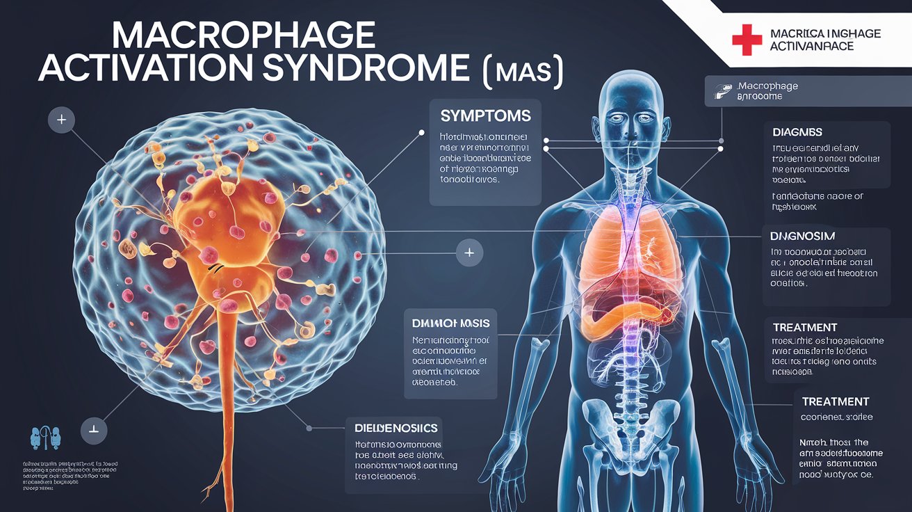
1. Introduction
2. What is Macrophage Activation Syndrome (MAS)?
3. What Triggers MAS?
4. Symptoms of Macrophage Activation Syndrome (MAS)
Blood Cell Line Depression: There is profound depression of all three blood cell lines, leading to:
Coagulation Abnormalities: An abnormal coagulation profile is common, which may include:
Pulmonary Complications: Some patients may develop pulmonary arterial hypertension, a serious complication associated with MAS.
Paradoxical Improvement in Underlying Disease: Interestingly, some patients may show a temporary improvement in their underlying inflammatory disease at the onset of MAS, with a decrease in arthritis symptoms and a drop in erythrocyte sedimentation rate (ESR).
Hyperferritinemia: Very high levels of ferritin in the blood are a significant laboratory hallmark of MAS, often exceeding 10,000 ng/mL during the acute phase.
5. How is MAS Diagnosed?
6. Complications of MAS

7. Treatment Options for MAS
Corticosteroid Therapy
Cyclosporine A Therapy
Etoposide Therapy
This protocol consists of:
Precautions with Etoposide:
Antithymocyte Globulin (ATG) Therapy
Biological Medications
Intravenous Immunoglobulin (IVIG) Therapy
8. Managing Macrophage Activation Syndrome (MAS) in Adults
Diagnosis:
Initial Treatment:
Combination Therapy:
Monitoring and Follow-Up:
Challenges in Management:
9. Living with MAS
10. Recent Advances in MAS Research
11. Conclusion
12. Frequently Asked Questions (FAQs)
1. Is MAS curable?
2. What is the survival rate for MAS?
3. Can MAS recur?
4. How can MAS be prevented?
5. Are there natural treatments for MAS?
6. What is the difference between MAS and HLH?
Recent Posts
Sequence Alignment plays a vital role in the subsequent analysis of NGS data, where millions of sequenced DNA fragments (reads) need to be aligned with a chosen reference sequence in a timely manner.
FASTQ files serve as the “raw data files” for any sequencing application, indicating that they are “unaltered.” Consequently, this file format is utilized for performing Quality Checks on sequencing reads. The Quality Check process is typically carried out using the FastQC tool developed by Simon Andrews from Babraham Bioinformatics.
FASTA format is a text-oriented format utilized for depicting either nucleotide sequences or peptide sequences, where nucleotides or amino acids are denoted by a single-letter code.
Mammalian expression systems enable the production of complex, functional recombinant proteins with proper folding and post-translational modifications. These systems are ideal for studying human proteins in a near-native environment, offering advantages in scalability, gene delivery, and purification. HEK293 and CHO cells remain the most widely used hosts, supporting both transient and stable expression strategies for academic and pharmaceutical applications.
Gas Chromatography (GC) stands as one of the most powerful and versatile analytical techniques used to separate and analyze compounds in complex mixtures. At its core, GC enables the identification and quantification of chemical substances based on their molecular composition and retention behaviors during migration through a chromatographic column.